Home » Without Label » Upper Thigh Anatomy / Lower Extremities Arteries And Nerves Anatomy Branches Kenhub / Related posts of muscle anatomy of upper thigh human muscle anatomy.
Upper Thigh Anatomy / Lower Extremities Arteries And Nerves Anatomy Branches Kenhub / Related posts of muscle anatomy of upper thigh human muscle anatomy.
Upper Thigh Anatomy / Lower Extremities Arteries And Nerves Anatomy Branches Kenhub / Related posts of muscle anatomy of upper thigh human muscle anatomy.. The posterior upper leg muscles provide your knees with mobility (extension, flexion and rotation) and strength. Thigh the thigh bears much of the load of the body's weight when a person is upright. Benjamin ma, md, professor, chief, sports medicine and shoulder service, ucsf department of orthopaedic surgery, san francisco, ca. It's the area that runs from the hip to the knee in each leg. The thigh muscles are divided into three compartments:
It contains many muscles and nerves but only has one bone, the femur, which is the longest and strongest bone. Upper leg anatomy and function the upper leg is often called the thigh. Muscles play an important role in the. The muscles that make up the quadriceps are the strongest and leanest of all muscles in the body. Rectus femoris muscle, one of the quadriceps muscles on the front of your thigh.
Ligaments Tendons And Muscles Of The Hip Joint Naples Best Hip Surgeon from zehrcenter.com The rectus femoris is located in the center of the thigh, while the vastus medialis is in the middle of the said body part. The four muscles all extend the lower leg. Also, we have prepared a special quiz for you to solidify your knowledge about the upper limb anatomy. People who play soccer have these specific muscles of the leg very well defined, so they're like a walking anatomy atlas for thigh muscles. The thigh bone, or femur, is the large upper leg bone that connects the lower leg bones (knee joint) to the pelvic bone (hip joint). Benjamin ma, md, professor, chief, sports medicine and shoulder service, ucsf department of orthopaedic surgery, san francisco, ca. Like the forearm, the upper leg, or thigh, has a dense arrangement of many muscles. Medial muscles adduct and rotate your thigh, and posterior flex your leg and extend your thigh.
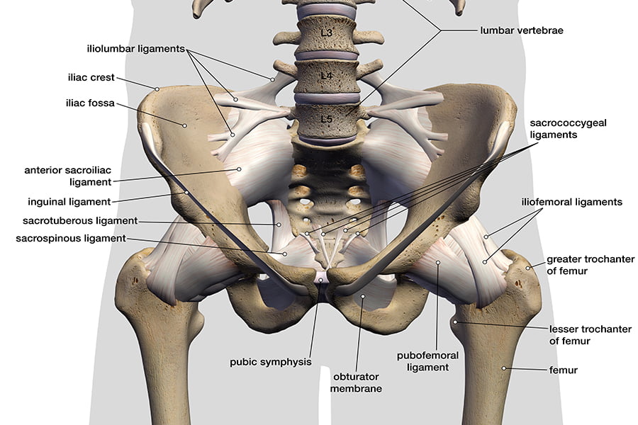
They have a lot to do with how your hips move.
Your leg muscles are some of the hardest working muscles in your body. Abductors are located on the upper portion of the outside of your thighs and hips, anchoring above on the pelvis, and below at various points on your outside thigh. Thigh the thigh bears much of the load of the body's weight when a person is upright. Anterior muscles extend your legs and flex your thighs. The thigh bone, or femur, is the large upper leg bone that connects the lower leg bones (knee joint) to the pelvic bone (hip joint). It's the area that runs from the hip to the knee in each leg. Rectus femoris muscle, one of the quadriceps muscles on the front of your thigh. People who play soccer have these specific muscles of the leg very well defined, so they're like a walking anatomy atlas for thigh muscles. Anatomynote.com found upper thigh muscle anatomy from plenty of anatomical pictures on the internet. Muscles play an important role in the. The largest muscle masses in the leg are present in the thigh and the calf. Pain in your calf or thigh can be caused by muscle cramps, a pulled or strained muscle, or issues related to your nerves. Find out everything about shoulder anatomy through our fun and engaging educational content.
Muscles play an important role in the. Ebraheim's educational animated video describes muscle anatomy of the thigh. The hamstring portion of the adductor magnus has a similar action to these muscles, but is located in the medial thigh. You can click the image to magnify if you cannot see clearly. It contains many muscles and nerves but only has one bone, the femur, which is the longest and strongest bone.
The Class Book Of Anatomy Designed For Schools Explanatory Of The First Principles Of Human Mechanism As The Basis Of Physical Education The Blood Vessels The Nervethat Supplies The Fore Part from c8.alamy.com The posterior upper leg muscles provide your knees with mobility (extension, flexion and rotation) and strength. This image added by admin. The muscles that make up the quadriceps are the strongest and leanest of all muscles in the body. Upper leg anatomy and function the upper leg is often called the thigh. Take the upper extremity anatomy quiz and learn more about the bones, joints, muscles and vessels of the upper extremity! Your leg muscles are some of the hardest working muscles in your body. Find out everything about shoulder anatomy through our fun and engaging educational content. Like the forearm, the upper leg, or thigh, has a dense arrangement of many muscles.
Iliopsoas muscle, a hip flexor muscle that attaches to the upper thigh bone.
The muscles that make up the quadriceps are the strongest and leanest of all muscles in the body. Thigh the thigh bears much of the load of the body's weight when a person is upright. In clinical anatomy the thigh muscles are divided into three groups: Medial muscles adduct and rotate your thigh, and posterior flex your leg and extend your thigh. The hamstring portion of the adductor magnus has a similar action to these muscles, but is located in the medial thigh. Ebraheim's educational animated video describes muscle anatomy of the thigh. Your leg muscles are some of the hardest working muscles in your body. Meanwhile, the vastus lateralis is on the side of the thigh, while the vastus intermedius is hidden below the rectus femoris(5). Anatomynote.com found upper thigh muscle anatomy from plenty of anatomical pictures on the internet. The muscles located within the posterior compartment of the thigh are the biceps femoris, semitendinosus and semimembranosus. Also, we have prepared a special quiz for you to solidify your knowledge about the upper limb anatomy. Muscles play an important role in the. Like the adductors, the abductors are also responsible for stabilizing your knees during athletic and everyday movement.
Like the adductors, the abductors are also responsible for stabilizing your knees during athletic and everyday movement. In clinical anatomy the thigh muscles are divided into three groups: It's the area that runs from the hip to the knee in each leg. The thigh muscles don't just move your legs. Muscles play an important role in the.
Legs To Base Line Connection Rectus Femoris Gluteus Maximus from www.baselinehealing.com Upper leg anatomy and function the upper leg is often called the thigh. People who play soccer have these specific muscles of the leg very well defined, so they're like a walking anatomy atlas for thigh muscles. Medial muscles adduct and rotate your thigh, and posterior flex your leg and extend your thigh. Find out everything about shoulder anatomy through our fun and engaging educational content. On the anterior side, the most prominent of the muscles are the sartorius muscle and the four muscles that make up quadriceps muscle group (the quads.) The thigh muscles are divided into three compartments: The posterior upper leg muscles provide your knees with mobility (extension, flexion and rotation) and strength. The rectus femoris is located in the center of the thigh, while the vastus medialis is in the middle of the said body part.
People who play soccer have these specific muscles of the leg very well defined, so they're like a walking anatomy atlas for thigh muscles.
Review date 7/8/2020 updated by: Upper leg anatomy and function the upper leg is often called the thigh. The hamstring portion of the adductor magnus has a similar action to these muscles, but is located in the medial thigh. People who play soccer have these specific muscles of the leg very well defined, so they're like a walking anatomy atlas for thigh muscles. The largest muscle masses in the leg are present in the thigh and the calf. Teachme anatomy part of the teachme series the medical information on this site is provided as an information resource only, and is not to be used or relied on for any diagnostic or treatment purposes. The thigh muscles are divided into three compartments: Find out everything about shoulder anatomy through our fun and engaging educational content. Pain in your calf or thigh can be caused by muscle cramps, a pulled or strained muscle, or issues related to your nerves. The thigh is the region between the hip and knee joints. The rectus femoris is located in the center of the thigh, while the vastus medialis is in the middle of the said body part. Thigh the thigh bears much of the load of the body's weight when a person is upright. We think this is the most useful anatomy picture that you need.